
Intro
Discover 5 key medical graphy terms, including radiography, mammography, and tomography, to understand medical imaging techniques, diagnostic procedures, and therapeutic applications.
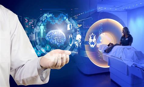
Medical imaging has become an indispensable tool in the field of medicine, allowing healthcare professionals to visualize the inside of the body and diagnose a wide range of medical conditions. With the advancement of technology, medical imaging has evolved to include various modalities, each with its unique characteristics and applications. In this article, we will delve into the world of medical imaging, exploring five key terms that are essential for understanding this complex and fascinating field.
The importance of medical imaging cannot be overstated, as it has revolutionized the way healthcare professionals diagnose and treat medical conditions. From X-rays to MRI scans, medical imaging has enabled doctors to non-invasively visualize the internal structures of the body, reducing the need for surgical interventions and improving patient outcomes. Moreover, medical imaging has also facilitated the development of personalized medicine, allowing healthcare professionals to tailor treatment plans to individual patients' needs.
As medical imaging continues to evolve, it is essential to stay up-to-date with the latest developments and advancements in the field. Whether you are a healthcare professional, a medical student, or simply someone interested in learning more about medical imaging, this article aims to provide a comprehensive overview of five key medical graphy terms. By understanding these terms, you will gain a deeper appreciation for the complexities and nuances of medical imaging, as well as its potential to transform the field of medicine.
Introduction to Medical Graphy

Types of Medical Graphy
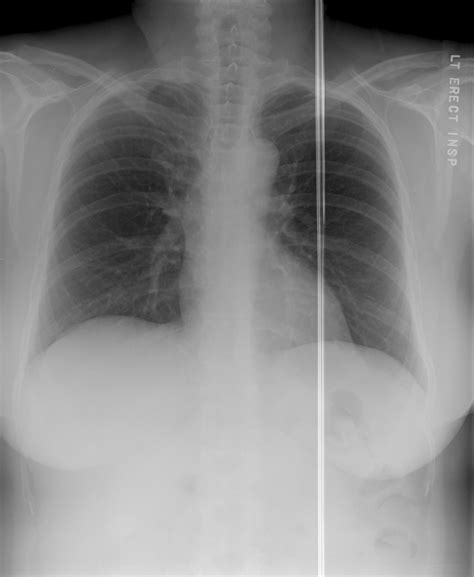
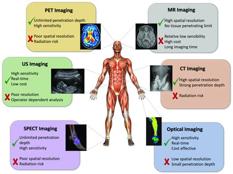
There are several types of medical graphy, each with its unique characteristics and applications. These include: * Radiography: This type of medical graphy uses X-rays to produce images of the body's internal structures. * Computed Tomography (CT) scans: This type of medical graphy uses a combination of X-rays and computer technology to produce cross-sectional images of the body. * Magnetic Resonance Imaging (MRI) scans: This type of medical graphy uses a strong magnetic field and radio waves to produce detailed images of the body's internal structures. * Positron Emission Tomography (PET) scans: This type of medical graphy uses a small amount of radioactive material to produce images of the body's metabolic activity. * Ultrasound: This type of medical graphy uses high-frequency sound waves to produce images of the body's internal structures.Term 1: Radiography

Applications of Radiography
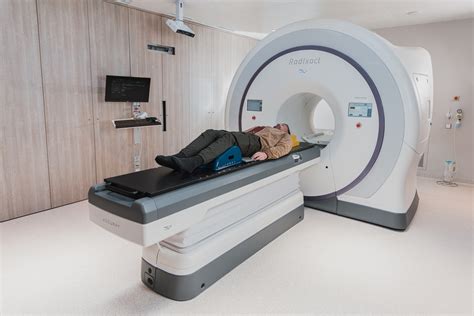
Radiography has a wide range of applications in medicine, including: * Diagnosing bone fractures and other musculoskeletal conditions * Detecting lung diseases, such as pneumonia and tuberculosis * Visualizing the digestive system, including the esophagus, stomach, and intestines * Guiding medical procedures, such as biopsies and surgeriesTerm 2: Computed Tomography (CT) Scans
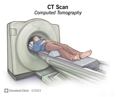
Applications of CT Scans
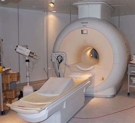
CT scans have a wide range of applications in medicine, including: * Diagnosing cancers, such as lung, liver, and pancreatic cancer * Detecting vascular diseases, such as atherosclerosis and aneurysms * Visualizing the brain and spinal cord, including the diagnosis of stroke and spinal cord injuries * Guiding medical procedures, such as biopsies and surgeriesTerm 3: Magnetic Resonance Imaging (MRI) Scans

Applications of MRI Scans
MRI scans have a wide range of applications in medicine, including: * Diagnosing neurological disorders, such as multiple sclerosis and Parkinson's disease * Detecting musculoskeletal conditions, such as tendonitis and ligament sprains * Visualizing the brain and spinal cord, including the diagnosis of stroke and spinal cord injuries * Guiding medical procedures, such as biopsies and surgeriesTerm 4: Positron Emission Tomography (PET) Scans
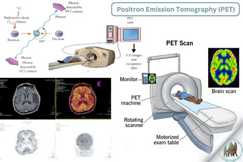
Applications of PET Scans
PET scans have a wide range of applications in medicine, including: * Diagnosing cancers, such as lung, breast, and colon cancer * Detecting neurological disorders, such as Alzheimer's disease and Parkinson's disease * Visualizing the heart and blood vessels, including the diagnosis of coronary artery disease * Guiding medical procedures, such as biopsies and surgeriesTerm 5: Ultrasound

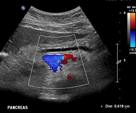
Applications of Ultrasound
Ultrasound has a wide range of applications in medicine, including: * Diagnosing obstetric and gynecological conditions, such as pregnancy and ovarian cysts * Detecting musculoskeletal conditions, such as tendonitis and ligament sprains * Visualizing the blood vessels, including the diagnosis of vascular diseases * Guiding medical procedures, such as biopsies and surgeriesMedical Imaging Gallery

In conclusion, medical imaging is a vital tool in the field of medicine, enabling healthcare professionals to diagnose and treat a wide range of medical conditions. By understanding the five key medical graphy terms discussed in this article, you will gain a deeper appreciation for the complexities and nuances of medical imaging, as well as its potential to transform the field of medicine. We invite you to share your thoughts and experiences with medical imaging in the comments section below, and to explore the many resources available online to learn more about this fascinating field. Whether you are a healthcare professional, a medical student, or simply someone interested in learning more about medical imaging, we hope that this article has provided a comprehensive and informative overview of the topic.